
Heart in Hand, Henry Ford Health System Leads the Way in Cardiac 3D Printing

 While we’re a long ways off yet from 3D printing organs for transplant, today’s reality is seeing 3D printed hearts saving patient lives in plastic. The structural heart team at Henry Ford Health System (HFHS) in Detroit is paving the way for those who will come after therm — and there will be many who follow where they are leading. I try to stay away from asserting my own predictions in the fast-growing, fast-changing world that is the 3D printing industry, but here I have no qualms; we will be seeing more hospitals setting up 3D printing labs.
The reality is that 3D printing as it is today isn’t yet viable for some of the big applications where industry will eventually be capitalizing on additive manufacturing as part of day-to-day operations. At the same time, however, what technology we do have immediately available has already proven to be enough to save lives when in the right hands. It doesn’t have to be the newest, the fastest, or the most expensive, either; while the financial outlay set out at HFHS was by no means insignificant, as the Henry Ford Innovation Institute (HFII) houses 3D printers from Stratasys, Formlabs, and MakerBot, none of these technologies are break-the-bank million-dollar machines. With these 3D printers — the Objet30 Pro being the workhorse and the Form 2 being increasingly put to use — the small, dedicated HFHS team has already touched more than 500 lives.

Two aspects of the work being done at HFHS highlight the ways in which the hospital system is setting itself up as a leader in the field, as saving lives is not a selfish endeavor; those working with advanced technology in Detroit want to see these processes expand well beyond their usage. While 500 heart procedures is by no means insignificant, that number can expand exponentially with the right systems in place to help adoption increase. Through seeking to commercialize the processes and creating a large physical library of patient heart models, the team at HFHS is looking to lead the way.
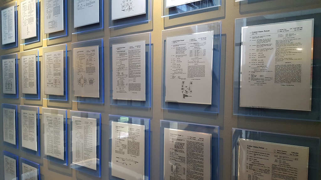
 Eric Myers, Product Designer, HFII, told me during my visit to Detroit early this month that, in fact, commercialization is a main goal for the institute. The staircase at HFII is a showroom of what he meant; the walls are covered with dozens of patents awarded to the institute. Surgical instruments, applications, and other forms of intellectual property from employees are being taken outside this environment through a push toward commercialization that can bring new technologies with proven success to a broader usage base. Specifically regarding 3D printed hearts, the team at HFHS has so far applied for three patents — with one of those granted so far, and licensed to Materialise — and published about half a dozen peer-reviewed research papers.
“Beyond touching our patients’ lives, we have had some commercial success,” Myers told me.
Dr. Dee Dee Wang, Director of Structural Heart Imaging at Henry Ford Hospital’s Center for Structural Heart Disease, expanded on this idea from a physician’s perspective. Along with the well-known gap between academic research and commercialization, often referred to as a valley of death, is the divide between research emerging from pioneering hospital settings and commercial availability.
“There’s a huge gap between lab science and what’s available commercially. Bridging that gap would be tremendous for patient care,” Dr. Wang explained.

[Image provided by HFHS]
“About 10 to 15 years are required” for this technology to take a stronger hold in cardiac centers, Dr. Wang said. “Most of our patients don’t have that.”

[Image provided by HFHS]
“This is not theoretical. We put this into action all the time,” Dr. William O’Neill, Medical Director, Henry Ford Center for Structural Heart Disease, told me.
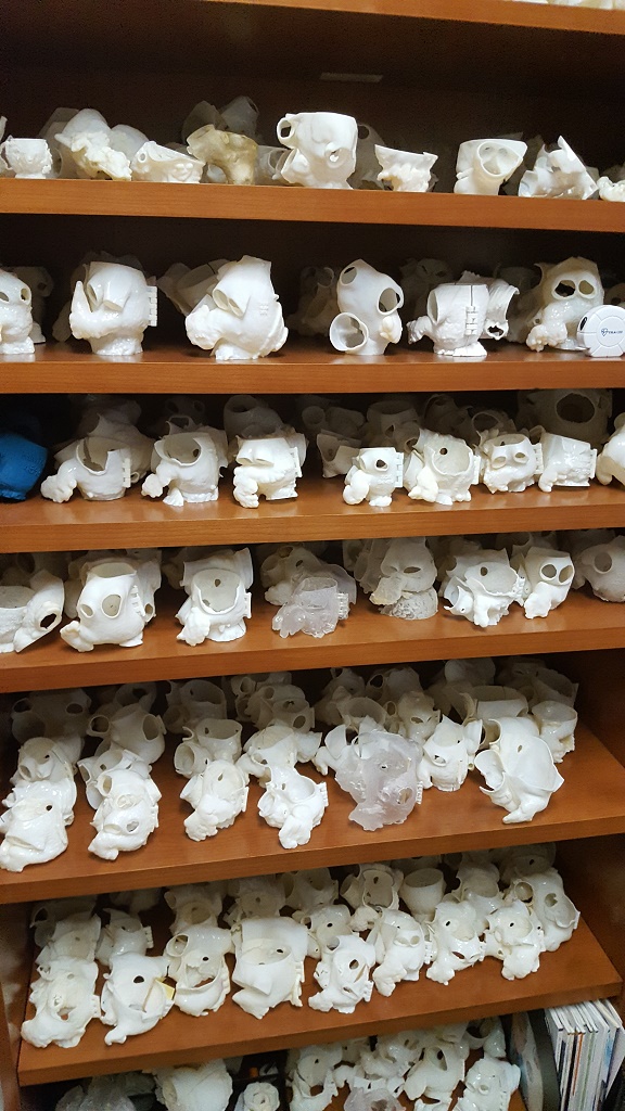
Demonstrating the volume of cases where 3D printing has already been applicable in their work is a large physical library. Reference libraries are an invaluable resource to medical professionals looking to gain more insight into current and future patient cases based on similar cases seen before. As 3D technologies come further into play in the medical field, we’ve seen that a library of 3D heart models and a 40-plus model physical library have proven to be of great benefit in Illinois; on the shelves in Dr. Wang’s office are several hundred 3D printed hearts.
-

- 3D printed hearts in Dr. Wang’s office
-

- Dr. Wang (L) and Marianne Rollet, Imaging/Applications Specialist

“This is something healthcare has never had before, and we don’t even know now how valuable this will be,” Myers told me of the untapped potential for value to be gained from this large, and growing, collection.
“Right now, information is only saved for six months, or you need to request it from the archives. With screen real estate, you could only look at two to three at a time. Now, [Dr. Wang] can walk to her shelf and pull five or six models to look at, and immediately see if the new patient has a similar case. She can compare it right away to other cases where we’ve seen success.”

Here I am holding a hinged 3D printed heart model at HFII
Having that information immediately at hand cuts back on both red tape and time involved in exploring commonalities to other cases the experienced team has faced. As we’ve heard from HFHS, their use of 3D printing has enhanced procedural time and success rates in complex cardiac cases; drawing from a growing pool of knowledge allows for a greater, and faster, comparison of cases and understanding of procedural enhancements.
As 3D printing at HFHS continues to expand, repercussions will ripple out from Detroit and potentially impact a much larger patient pool. Highly accurate 3D printed models of patients’ hearts offer benefits in training, procedural preparation, patient education, and future understanding and case comparison.
[All photos unless otherwise credited: Sarah Goehrke]
Subscribe to Our Email Newsletter
Stay up-to-date on all the latest news from the 3D printing industry and receive information and offers from third party vendors.
You May Also Like
Precision at the Microscale: UK Researchers Advance Medical Devices with BMF’s 3D Printing Tech
University of Nottingham researchers are using Boston Micro Fabrication‘s (BMF) 3D printing technology to develop medical devices that improve compatibility with human tissue. Funded by a UK grant, this project...
3D Printing Webinar and Event Roundup: April 21, 2024
It’s another busy week of webinars and events, starting with Hannover Messe in Germany and continuing with Metalcasting Congress, Chinaplas, TechBlick’s Innovation Festival, and more. Stratasys continues its advanced training...
3D Printing Webinar and Event Roundup: March 17, 2024
It’s another busy week of webinars and events, including SALMED 2024 and AM Forum in Berlin. Stratasys continues its in-person training and is offering two webinars, ASTM is holding a...
3D Printed Micro Antenna is 15% Smaller and 6X Lighter
Horizon Microtechnologies has achieved success in creating a high-frequency D-Band horn antenna through micro 3D printing. However, this achievement did not rely solely on 3D printing; it involved a combination...


